
CT Scan Services
SR Scanning Center offers advanced 80 Slice CT Scanning technology for precise and rapid diagnostics.
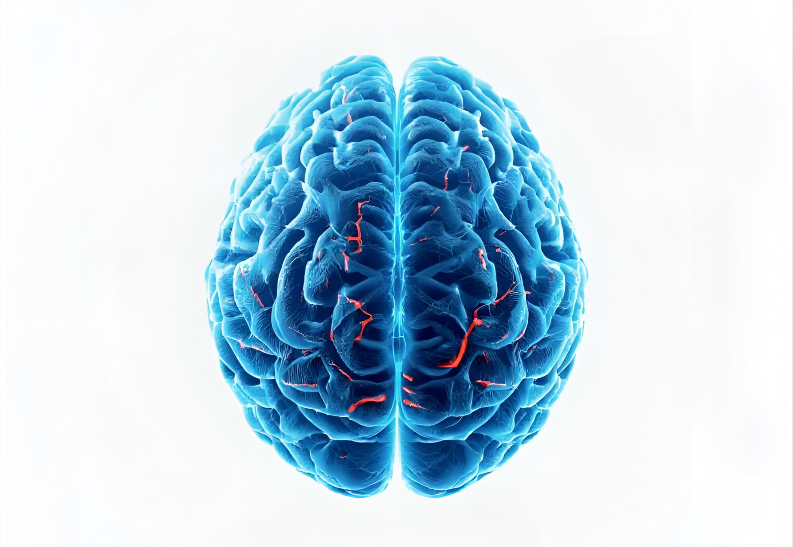
CT Brain (Plain / Contrast)
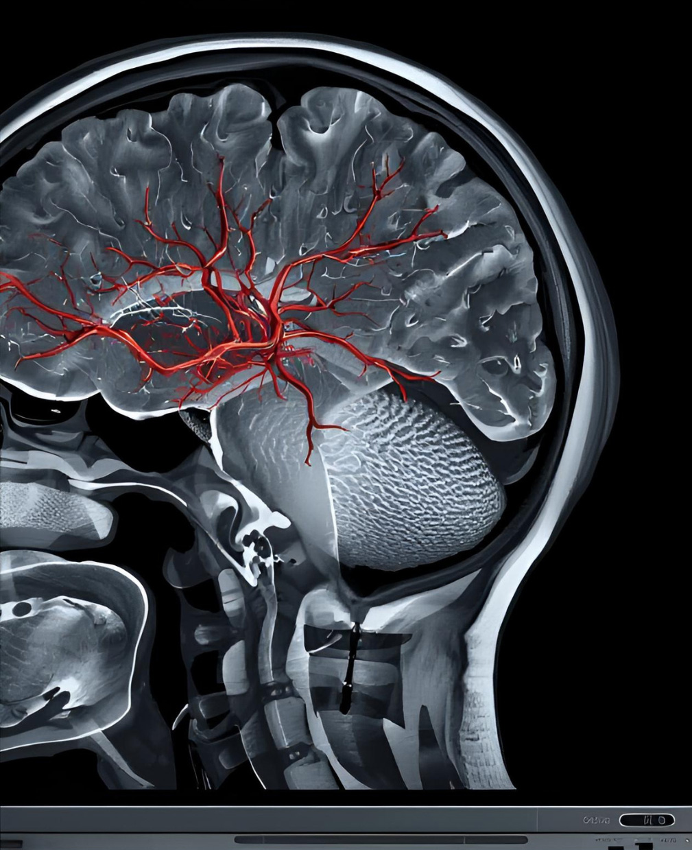
Our state-of-the-art 80 Slice CT scanner offers unparalleled speed and resolution for brain imaging. A CT scan of the brain is a non-invasive diagnostic imaging procedure that uses special X-ray equipment to produce horizontal, or axial, images (often called slices) of the head. These images provide detailed information about brain tissue and brain structures, including arteries and veins, which are not visible on standard X-rays. It is the modality of choice for acute neurological emergencies.
This scan is the first line of defense in emergency medicine for detecting strokes, head injuries, and intracranial bleeding. The speed of our 80-slice machine means the scan is completed in mere seconds, reducing the need for patients to hold still for long periods, which is crucial in trauma cases where time is of the essence. It is also highly effective in detecting brain tumors, hydrocephalus, and structural anomalies that may cause chronic symptoms.
For patients suffering from chronic headaches, dizziness, or seizures, a CT Brain scan helps rule out serious underlying conditions. Depending on the clinical requirement, contrast dye may be injected to enhance the visibility of tumors or infection, allowing our radiologists to provide a precise diagnosis. The advanced reconstruction capabilities allow us to see the brain in 3D, aiding surgeons in planning complex procedures.
Key Highlights
- Rapid detection of stroke and hemorrhage (bleeding).
- High-resolution imaging for trauma and accident cases.
- Non-invasive evaluation of chronic headaches and seizures.
- 3D reconstruction for surgical planning and tumor assessment.
FAQs - Brain CT
For a plain CT scan of the brain, no fasting is required, and you can eat and drink normally. However, if your doctor has ordered a contrast-enhanced scan (CECT), you will be required to fast for at least 4 hours prior to the appointment to prevent nausea.
The radiation dose is low and considered safe for diagnostic purposes. Our 80-slice scanner uses advanced technology to minimize exposure while maintaining image quality. The benefits of an accurate diagnosis in emergency situations far outweigh the minimal risk.
The actual scanning process is incredibly fast, often taking less than a minute. However, the total time in the room for positioning and preparation may take about 10-15 minutes. It is a very quick and painless procedure.
CT Orbits
A CT scan of the orbits provides detailed images of the eye sockets (orbits), eyes, and surrounding bone and soft tissues. This scan is essential for diagnosing trauma to the eye area, including fractures of the orbital bones (blowout fractures) which can trap eye muscles and affect vision. Our high-resolution scanner ensures that even minute fractures or foreign bodies are detected with precision.
Beyond trauma, CT Orbits are used to investigate protrusions of the eye (proptosis), infections (orbital cellulitis), and tumors such as optic nerve gliomas or lymphomas. It allows for the visualization of the optic nerve and the muscles that control eye movement, helping doctors determine the mechanical or neurological cause of double vision or sudden vision loss.
The procedure is quick and painless. We use specific protocols to reduce radiation exposure to the sensitive lens of the eye while maintaining image quality high enough to guide surgical interventions if necessary. It is far superior to standard X-rays for evaluating the complex bony anatomy of the face and eye sockets.
Key Highlights
- Detailed visualization of orbital fractures and foreign bodies.
- Assessment of orbital tumors and infections (cellulitis).
- Evaluation of the optic nerve and eye muscles.
- Critical for planning corrective eye or facial surgeries.
FAQs - Orbits
No, you should remove contact lenses before the scan to avoid artifacts in the images. We also recommend removing any eye makeup, especially mascara, which might contain metallic particles that can interfere with the image quality.
Contrast injection is typically only used if the doctor suspects an infection, inflammation, or a tumor within the orbit. For routine trauma cases involving fractures, a plain scan without contrast is usually sufficient.
Yes, the CT scan does not affect your vision or ability to drive. Unless you were given a sedative (which is rare for this procedure), you can resume normal activities immediately after leaving the clinic.
CT Paranasal Sinuses (PNS)
CT of the Paranasal Sinuses (PNS) is the gold standard for evaluating chronic sinusitis. It provides a clear roadmap of the air-filled cavities surrounding the nose, allowing ENT surgeons to see blockages, polyps, or thickening of the sinus lining that cannot be seen with a simple nasal endoscopy. It is invaluable for patients suffering from chronic congestion or sinus pressure.
This scan is critical for planning FESS (Functional Endoscopic Sinus Surgery). Our 80 Slice scanner creates multi-planar reconstructions (Coronal, Sagittal, and Axial views) which help in identifying anatomical variations like a deviated septum or concha bullosa that might be contributing to sinus headaches and breathing issues.
It is also used to assess fungal infections, tumors of the nasal cavity, and trauma to the facial region. The scan is extremely fast, usually taking less than 20 seconds of actual exposure time, making it easy even for claustrophobic patients.
Key Highlights
- Gold standard for diagnosing Chronic Sinusitis.
- Essential for FESS (Endoscopic Sinus Surgery) planning.
- Detects nasal polyps, deviated septum, and fungal infections.
- Multi-planar views provide a complete 3D roadmap.
FAQs - PNS
No, the CT PNS scan is completely painless and non-invasive. Nothing touches your face; you simply lie still on the table while the scanner ring moves around your head.
No, you can continue your medications unless specifically told otherwise by your doctor. In fact, knowing the current state of your sinuses while on medication can sometimes help the doctor evaluate treatment effectiveness.
CT provides a 3D view without overlapping bones, unlike X-rays which are 2D and often obscure deep sinus cavities. CT is far more sensitive and specific for sinus disease than a standard X-ray.
CT Neck (Soft Tissue)
A CT scan of the neck focuses on the soft tissues, including the throat, tonsils, thyroid gland, and trachea. It is frequently ordered to investigate lumps or masses in the neck, swollen lymph nodes, or persistent hoarseness. The high-speed capture helps minimize artifacts from swallowing or breathing, providing clear images of this complex area.
This scan is vital for staging cancers of the head and neck, allowing oncologists to see if the disease has spread to lymph nodes or surrounding structures. It helps in distinguishing between cysts, infections (abscesses), and solid tumors. Contrast dye is almost always used to differentiate blood vessels from lymph nodes and masses.
Additionally, it can evaluate the salivary glands for stones or infections and check the airway for narrowing or obstructions. It is a comprehensive tool for evaluating any unexplained pain or swelling in the neck region.
Key Highlights
- Evaluates thyroid nodules, lymph nodes, and neck masses.
- Crucial for staging head and neck cancers.
- Detects abscesses and salivary gland stones.
- Fast imaging minimizes motion blur from swallowing.
FAQs - Neck CT
No, the machine is open at both ends and creates plenty of space. You just lie flat on your back. If you feel anxious, let our technicians know, and they will help guide you through the process comfortably.
The neck is full of major blood vessels like the carotid artery. Contrast dye makes these vessels bright white, allowing the radiologist to easily distinguish them from lymph nodes and tumors, which look similar on plain scans.
You will be asked not to swallow for a few seconds while the images are being taken. Swallowing moves the larynx and can cause blurry images, so holding still is very important for a clear result.
CT Chest (Plain / Contrast)
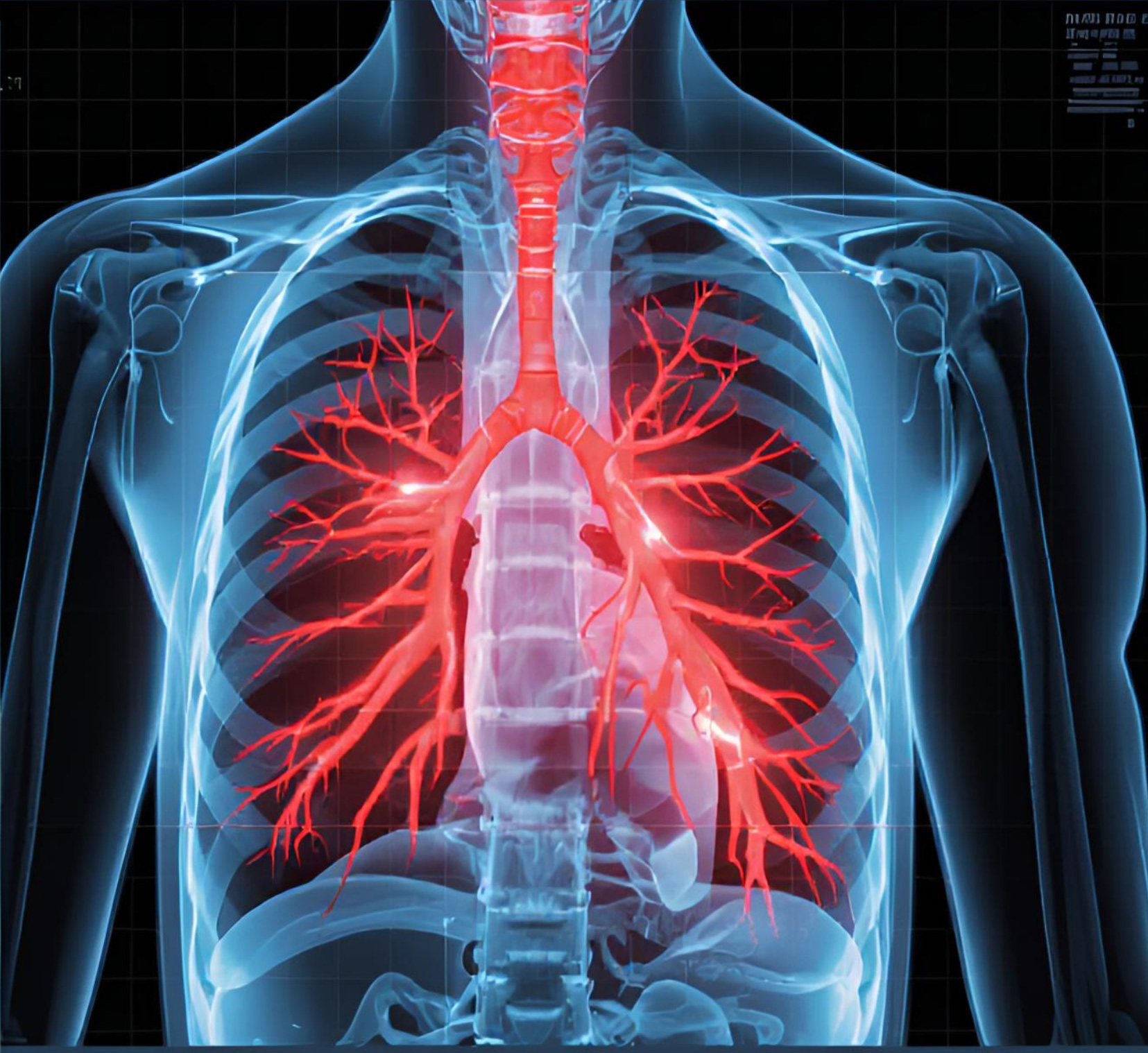
A routine CT Chest is used to diagnose conditions affecting the lungs, heart, and mediastinum. Unlike a standard chest X-ray, a CT scan provides detailed cross-sectional views that can identify small nodules, tumors, or fluid collections that X-rays might miss. It is the most effective tool for investigating chest pathology.
This scan is commonly used to investigate chest pain, shortness of breath, or chronic cough. It is essential for lung cancer screening in high-risk patients. When used with contrast (CECT Chest), it can highlight the blood vessels to rule out pulmonary embolism (a blood clot in the lung) or aortic aneurysms.
It also evaluates the chest wall, rib cage, and spine in cases of severe trauma, ensuring no internal bleeding or organ damage has occurred. Whether for infection, tumor, or trauma, the CT Chest provides a definitive diagnosis.
Key Highlights
- Detects lung nodules, tumors, and pneumonia early.
- Rules out Pulmonary Embolism (with contrast).
- Evaluates chest trauma and rib fractures.
- Gold standard for complicated chest infections.
FAQs - Chest CT
Yes, usually for 10-15 seconds. Breathing causes the lungs to move, which blurs the picture. Our technicians will coach you on breathing instructions before the scan begins.
Wear loose, comfortable clothing. You should avoid clothes with metal zippers, buttons, or underwire bras, as metal causes streaks on the images. We may provide a hospital gown if necessary.
The scan itself is very fast, typically completed in under a minute. However, if contrast dye is used, there may be a short wait while the IV is inserted and the dye circulates.
CT Facial Bones
A CT of the facial bones is primarily used to assess trauma. The face has a complex structure of many small, delicate bones. Our 80-slice scanner creates 3D reconstructions that are vital for maxillofacial surgeons to plan reconstructive surgeries after accidents or assaults. It reveals displacements and fractures that are invisible on standard X-rays.
Beyond trauma, this scan helps in planning surgeries for facial deformities or prior to dental implants to assess bone density and nerve location. It can also identify tumors, cysts within the jawbones, or infections spreading from the teeth to the sinus cavities.
The high resolution ensures that even hairline fractures in areas like the orbital floor or zygomatic arch are clearly visible. This leads to better cosmetic and functional outcomes for patients requiring surgery.
Key Highlights
- Essential for evaluating facial trauma and fractures.
- 3D imaging aids in reconstructive surgery planning.
- Assesses jaw tumors and dental pathology.
- High resolution for delicate facial structures.
FAQs - Facial Bones
Removable dentures must be taken out to avoid blocking the view. Fixed implants or braces can stay, though they may cause minor artifacts (streaks) which our software attempts to correct.
No, your head goes into the scanner, but the machine is wide and open, not a long tunnel. Most patients tolerate facial scans very well without anxiety.
None at all. You just need to lie very still for a few seconds. The procedure is entirely non-contact and non-invasive.
HRCT Chest (High Resolution)
High-Resolution CT (HRCT) uses specific scanning techniques to visualize the lung parenchyma (tissue) in extreme detail. It is the gold standard for diagnosing interstitial lung diseases (ILD), pulmonary fibrosis, and bronchiectasis. Unlike a standard CT, HRCT uses very thin slices (1mm or less) to capture the fine architecture of the lungs.
This scan became widely recognized during the COVID-19 pandemic for assessing the severity of viral pneumonia (CORADS scoring). It accurately measures the percentage of lung involvement and damage, helping doctors determine the need for oxygen therapy or hospitalization. It is far more sensitive than a chest X-ray.
HRCT is generally performed without contrast. It helps pulmonologists decide on treatments for chronic lung conditions and monitor whether a disease is progressing or responding to therapy. It effectively differentiates between active inflammation and permanent scarring.
Key Highlights
- Gold standard for Interstitial Lung Disease (ILD).
- Accurate scoring for COVID-19 and viral pneumonia.
- Detects Pulmonary Fibrosis and Bronchiectasis.
- Uses thin slices for maximum lung detail.
FAQs - HRCT
Typically, no contrast dye is needed for HRCT lung studies because air in the lungs provides natural contrast against the tissue. It is a simple "plain" scan.
HRCT takes thinner slices (1mm vs 5mm) and uses a sharper reconstruction algorithm to see the fine texture of lung tissue, which is necessary for diagnosing fibrosis.
Yes, HRCT is very effective at detecting active Tuberculosis lesions, cavities, or old TB scarring that might not be clearly visible on a regular chest X-ray.
CT Abdomen & Pelvis
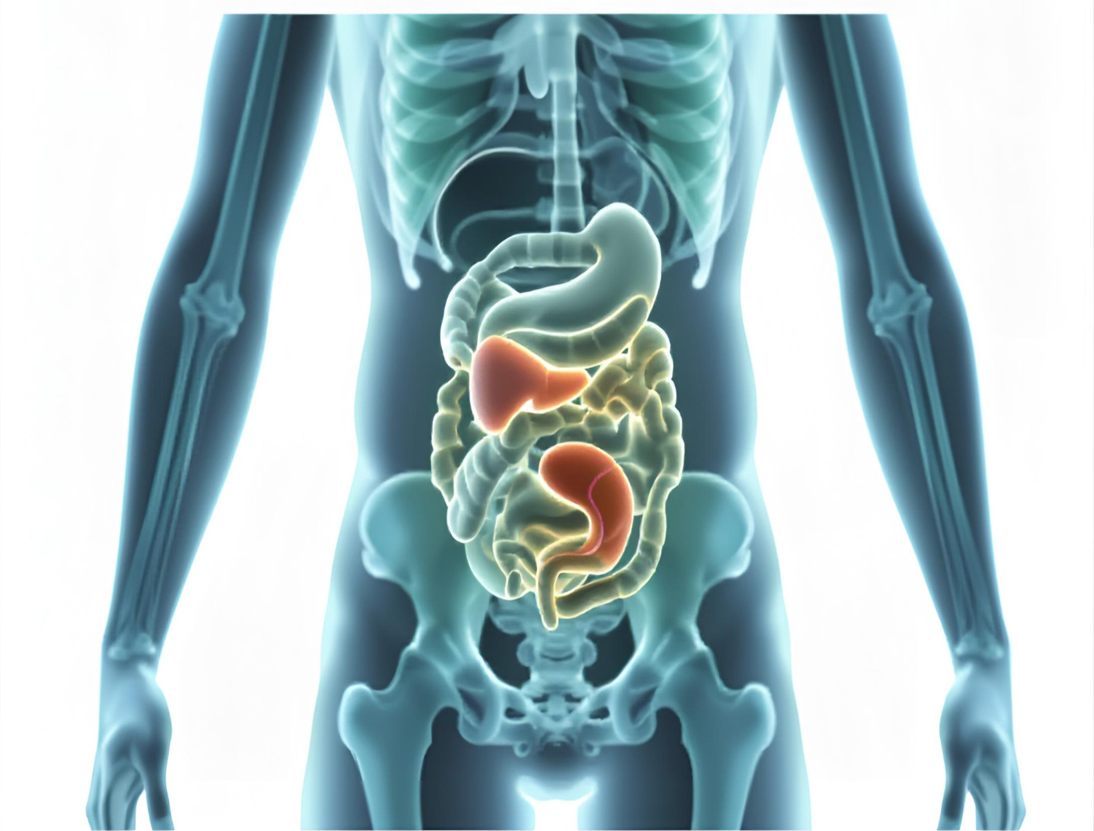
CT of the Abdomen and Pelvis provides a comprehensive view of the liver, spleen, pancreas, kidneys, intestines, and bladder. It is the preferred modality for diagnosing the cause of acute abdominal pain, such as appendicitis, diverticulitis, or pancreatitis. It allows doctors to see inside solid organs and bowel loops simultaneously.
For cancer patients, this scan is commonly used for staging to see if tumors have spread (metastasized) to the liver or lymph nodes. We often use both oral contrast (which you drink to highlight the stomach and intestines) and IV contrast (injected to highlight organs and blood vessels) for maximum diagnostic accuracy.
It is also used to assess abdominal trauma after accidents, ensuring there is no internal bleeding or organ rupture. The speed of our scanner minimizes motion blur from breathing or bowel movements, ensuring crisp images.
Key Highlights
- Diagnoses Appendicitis, Pancreatitis, and Diverticulitis.
- Staging and monitoring of abdominal cancers.
- Evaluates trauma for internal bleeding or organ damage.
- Uses Oral and IV contrast for comprehensive imaging.
FAQs - Abdomen
Drinking contrast or water helps expand the stomach and bowel loops. This ensures the radiologist doesn't mistake a collapsed bowel for a tumor or mass.
You generally need to fast for 4-6 hours before an abdominal CT with contrast. An empty stomach reduces the risk of nausea when the contrast dye is injected.
The contrast dye is cleared by the kidneys. We check your serum creatinine levels beforehand to ensure your kidney function is normal and can handle the dye safely.
CT Pelvis
While often combined with the abdomen scan, a dedicated CT Pelvis scan focuses on the reproductive organs, bladder, prostate (in men), and pelvic bones. In orthopedic cases, it is critical for analyzing complex hip fractures or planning hip replacement surgeries, providing 3D views of the pelvic girdle.
In urology and gynecology, it helps detect tumors of the bladder, uterus, or ovaries. While MRI or Ultrasound is sometimes preferred for soft tissue in this area, CT is superior for detecting calcifications, bone involvement, and enlarged lymph nodes in the pelvic region.
It is also used to investigate undiagnosed pelvic pain or deep abscesses. 3D reconstruction of the pelvic bones is standard protocol for trauma patients at our center to check for fractures.
Key Highlights
- Detailed imaging of pelvic fractures and hip joints.
- Evaluation of bladder, prostate, and reproductive organs.
- Detection of pelvic masses and abscesses.
- Often performed in conjunction with Abdomen CT.
FAQs - Pelvis
CT uses radiation and is generally NOT used to check for pregnancy as it can harm the fetus. Ultrasound is the safe method for pregnancy checks. If you might be pregnant, tell us immediately.
Sometimes a full bladder helps push the bowel out of the way and visualizes the bladder wall better. The technician will instruct you on whether to drink water before the scan.
The actual scan takes only a few seconds. The main time is spent positioning you correctly on the table to ensure the pelvis is centered.
CT KUB (Kidney, Ureter, Bladder)
CT KUB is the gold standard investigation for renal colic (severe kidney stone pain). Unlike ultrasound, which might miss stones lodged in the ureter, CT KUB visualizes the entire urinary tract from the kidneys down to the bladder. It accurately measures the size, location, and density (hardness) of the stone.
This detailed information helps urologists decide whether the stone is likely to pass on its own, requires lithotripsy (shock wave therapy), or needs surgical removal. It can also detect obstruction (hydronephrosis) where the kidney is swollen due to blockage, which can damage the kidney if left untreated.
Usually, this is a "Plain" scan, meaning no injection is required, making it quick and safe even for patients with compromised kidney function. It takes less than 5 minutes from entering the room to leaving.
Key Highlights
- Most accurate test for Kidney Stones (Renal Calculi).
- Detects ureteral blockage and Hydronephrosis.
- Determines stone size and hardness for treatment planning.
- Quick, non-invasive, and usually requires no contrast.
FAQs - KUB
For detecting stones, no contrast is needed. If the doctor suspects a tumor or complex cyst, they might order a contrast scan, but that is different from a standard KUB.
CT is incredibly sensitive and can detect stones as small as 1-2mm that would be completely invisible on an X-ray or Ultrasound.
Yes, significantly. X-rays can miss "radiolucent" stones (like uric acid stones), whereas CT sees all types of stones regardless of their composition.
CT Spine (Cervical / Dorsal / Lumbar)
CT Spine imaging is crucial for assessing the bony structures of the vertebral column. It is commonly performed for trauma patients to rule out fractures, dislocations, or bone fragments in the spinal canal following an accident. It provides far greater detail of the bone than an MRI scan.
It is also used to diagnose spinal stenosis (narrowing of the spinal canal), disc herniation (though MRI is superior for soft tissue, CT is used if the patient has a pacemaker or metal implants), and spinal tumors. It effectively shows arthritic changes and bone spurs.
Post-operative patients often get CT scans to check the precise placement of screws, rods, and cages used in spinal fusion surgeries. Our metal-artifact reduction software helps clear up images around surgical implants to ensure everything is healing correctly.
Key Highlights
- Superior imaging for spinal fractures and trauma.
- Assesses healing and hardware placement after surgery.
- Diagnoses spinal stenosis and bone spurs.
- Alternative to MRI for patients with pacemakers.
FAQs - Spine
CT is best for looking at bones and fractures. MRI is best for soft tissues like the spinal cord, nerves, and herniated discs. Your doctor chooses based on what they suspect.
Yes! Unlike MRI, which uses strong magnets that can stop a pacemaker, CT uses X-rays and is perfectly safe for patients with pacemakers or cochlear implants.
You lie on a hard table, which can be slightly uncomfortable if you have back pain, but the scan is very fast, so the discomfort is short-lived.
CT Urogram
A CT Urogram (CT IVU) is a specialized multiphasic exam used to evaluate the entire urinary tract (kidneys, ureters, and bladder) in great detail. It is primarily used to investigate blood in the urine (hematuria) to rule out cancers of the urinary system (Transitional Cell Carcinoma).
This procedure involves taking images before contrast, and then at specific time intervals after contrast injection. This allows us to see how well the kidneys filter the dye and excrete it into the ureters and bladder. It essentially functions as a functional and anatomical test combined.
It creates a 3D anatomical map of the urinary system, helping to identify strictures (narrowing), congenital anomalies (like horseshoe kidney), and tumors. It is far more detailed than a standard IVP X-ray study used in the past.
Key Highlights
- Investigates Hematuria (blood in urine).
- Detects cancers of the kidney lining and bladder.
- Evaluates kidney function and drainage.
- Replaces traditional IVP X-ray studies.
FAQs - Urogram
Because we take delayed images to watch the dye drain, the total time in the room may be 15-20 minutes, which is longer than a standard CT.
You will be asked to empty your bladder before the scan starts, but by the end of the scan, your bladder will fill with the contrast dye naturally.
Yes, 4 hours fasting is required because intravenous contrast dye is always used for a CT Urogram.
Virtual Bronchoscopy
Virtual Bronchoscopy is a non-invasive technique that uses CT data to create a 3D "fly-through" view of the airways (trachea and bronchi). It simulates the view a doctor would get from an actual invasive bronchoscopy scope but without inserting any tubes down the throat.
It is particularly useful for evaluating narrowing of the airway (stenosis), detecting foreign bodies, or assessing tumors that are blocking the air passages. Unlike a real scope, the "virtual" camera can pass through tight blockages to see what lies beyond the obstruction.
This is strictly a diagnostic tool performed via software processing of a high-resolution Chest CT. It requires no anesthesia, carries no risk of airway injury or bleeding, and is very comfortable for the patient.
Key Highlights
- Non-invasive view of the inside of airways.
- Navigates past strictures that block physical scopes.
- No anesthesia or sedation required.
- Useful for surgical planning of airway tumors.
FAQs - Virtual Bronchoscopy
No, this is a computer simulation based on a standard CT scan. Nothing touches your throat or mouth at all.
It is very accurate for viewing the structure, but unlike real bronchoscopy, it cannot take tissue samples (biopsy) or remove objects.
The scan takes seconds; the computer processing takes a bit longer, but you don't have to wait in the machine for that part.
CT Joints (Shoulder, Knee, Elbow, etc.)
CT scans of the joints are primarily used to evaluate complex fractures that involve the articular surface (the moving part of the joint). Standard X-rays often cannot show the alignment of shattered bone fragments (comminuted fractures), which is critical for surgical repair.
It is commonly used for the shoulder, elbow, wrist, hip, knee, and ankle. 3D Volume Rendering (VR) images allow orthopedic surgeons to "see" the fracture from all angles and plan exactly where to place screws and plates to restore movement.
CT is also used to diagnose loose bodies (bone chips) floating in the joint space, which can cause locking or pain. In some cases, it helps diagnose gout by detecting uric acid crystals deposited in the joints.
Key Highlights
- Essential for complex joint fractures.
- Detects loose bodies and bone fragments.
- Used for diagnosing Gout crystals.
- 3D planning for orthopedic surgery.
FAQs - Joints
It depends. For ligaments (ACL) and meniscus tears, MRI is better. For bone fractures and alignment, CT is better.
We try to make you comfortable, but keeping an injured joint still can be tricky. We use pillows and pads to support the limb.
The images are available quickly, but the full report with 3D analysis usually takes a few hours for the radiologist to prepare.
CT Extremities (Upper/Lower Limbs)
CT of the extremities covers the long bones (humerus, femur, tibia, etc.) and hands/feet. It is frequently used to detect occult fractures—hairline cracks in the bone that don't show up on initial X-rays but cause persistent pain. It provides a detailed look at bone integrity.
It is also vital for evaluating bone tumors (osteosarcoma) or deep infections (osteomyelitis). The scan determines how much bone has been destroyed and if the infection has formed a sequester (dead bone). This guides surgeons on how much tissue needs to be removed.
Our scan length measurement feature helps in evaluating limb length discrepancies in children or after trauma, ensuring accurate measurements for corrective shoes or surgery.
Key Highlights
- Detects hidden (occult) fractures missed by X-ray.
- Evaluates bone tumors and osteomyelitis.
- Accurate measurement of limb length discrepancy.
- Non-invasive assessment of bone healing.
FAQs - Extremities
Often, only the limb being scanned needs to be in the center of the machine. Your head and body might remain outside the ring, reducing claustrophobia.
Yes, CT X-rays can penetrate plaster or fiberglass casts easily, allowing doctors to check bone healing without removing the cast.
Only if we are looking for infection, abscess, or tumors. For simple fractures, plain scans are sufficient.
3D Reconstruction
3D Reconstruction is not a separate scan but a post-processing technique available with our 80-slice scanner. It takes the hundreds of 2D slices generated during a CT scan and stacks them digitally to create a photorealistic 3D model of the patient's anatomy.
This is invaluable for surgeons. For example, in complex facial fractures or crushed pelvis cases, the surgeon can rotate the 3D image on the screen to understand the exact position of every bone fragment before entering the operating room. It reduces surgical time and improves outcomes.
It is also used in angiography to show the "tree" of blood vessels, helping to visualize blocked arteries in 3D space relative to bones and organs. Patients also find 3D images much easier to understand than traditional black-and-white slices.
Key Highlights
- Photorealistic anatomical models.
- Critical for complex surgical planning.
- Better visualization of vascular trees and fractures.
- Easier for patients to understand their condition.
FAQs - 3D
It is usually included in the price of specific scans like trauma or angiograms where it is medically necessary. Ask our reception for details.
No, the 3D image is built from the data already captured during the initial scan. No extra radiation is required.
Yes, we can provide specific 3D images on the film or the full dataset on a CD/USB for your doctor to review.
CT Angiogram (CTA)
A CT Angiogram (CTA) is an advanced test that uses contrast dye and high-speed scanning to visualize blood vessels (arteries and veins) throughout the body. It is used to detect aneurysms (bulging vessels), stenosis (narrowing from plaque), and dissections (tears in the vessel wall).
Common types include Pulmonary Angiogram (for clots in lungs), Cerebral Angiogram (for brain aneurysms), Carotid Angiogram (neck arteries supplying brain), and Peripheral Angiogram (for blockages in legs causing pain). It provides a roadmap for vascular surgeons.
Because our 80-slice scanner is incredibly fast, it captures the image exactly when the dye passes through the arteries, providing crystal clear vascular maps without the need for invasive catheters used in traditional angiography.
Key Highlights
- Non-invasive imaging of arteries and veins.
- Detects aneurysms, blockages, and blood clots.
- Includes Pulmonary, Cerebral, and Peripheral Angio.
- Replaces invasive catheter angiography for diagnosis.
FAQs - Angiogram
Through a cannula (IV line) in your arm using a pressure injector that delivers the dye quickly to ensure it fills the arteries at the right moment.
You may feel a sudden warm flush spreading through your body or a metallic taste in your mouth. This is normal and passes within seconds.
Yes, provided your kidney function is normal (we check Serum Creatinine beforehand) and you have no history of severe allergy to iodine.
CT Temporal Bone / Inner Ear
This is a specialized high-resolution scan of the ear structures. It is primarily used to investigate hearing loss, tinnitus (ringing in the ears), and vertigo (dizziness). The temporal bone contains the tiny bones of the middle ear (ossicles) and the cochlea, which are clearly visible on this scan.
It is essential for evaluating chronic ear infections (CSOM) and Cholesteatoma (a destructive skin growth in the ear). Surgeons rely on this scan before performing mastoidectomies or cochlear implant surgeries to understand the patient's anatomy and avoid complications.
It is also used to assess fractures of the skull base after head trauma, which can cause leakage of brain fluid (CSF) from the ear. The sub-millimeter precision of our scanner is vital here.
Key Highlights
- Investigates Hearing Loss and Vertigo.
- Detects Cholesteatoma and Chronic Ear Infections.
- Pre-operative planning for Cochlear Implants.
- Assesses skull base fractures and CSF leaks.
FAQs - Temporal Bone
The machine makes a whirring sound, but it is much quieter than an MRI. You will just hear a gentle hum during the scan.
Usually no, unless the doctor is checking for a tumor like an Acoustic Neuroma or vascular issues. Most ear scans are plain.
It is very quick, typically 10-15 seconds scan time, though we take time to position your head perfectly straight.

Precision Imaging Meets Compassionate Care.
At SR Scanning Center, we combine advanced medical technology with a patient-first approach. Our 80 Slice CT Scanner delivers rapid, high-resolution images, ensuring that your diagnosis is both quick and accurate.
With over 90% of health decisions based on diagnostic results, we are committed to being the trusted healthcare partner you need for critical decisions.
Expert Radiologists
Our team consists of highly qualified radiologists and technicians dedicated to providing accurate reports and exceptional patient care.
Advanced Technology
Featuring the 80 Slice CT Scanner for sub-millimeter precision, reduced radiation dose, and faster scan times.
Process
Your Journey to a Diagnosis: Simple, Fast, and Reliable.
We understand that medical testing can be stressful. Our process is designed to be as smooth and efficient as possible. From booking your appointment to receiving your detailed report, our team is here to guide you every step of the way.
Most routine scans are completed within minutes, and our digital reporting system ensures your doctor gets the results they need without delay.
-
High-Speed Scanning
-
Minimal Radiation Dose
-
Digital & Print Reports
Schedule Scan
Call us or book online. Let us know if you have allergies, kidney issues, or if contrast is prescribed.
Contact UsArrive & Prep
Bring your doctor's prescription. If contrast is required, you may need to fast for 4 hours beforehand.
View Prep InfoThe Scan
Our expert technicians will guide you. The 80-slice scan is non-invasive and takes only a few seconds.
Learn MoreGet Results
Our radiologists analyze the images. You will receive high-quality films and a detailed report promptly.
View Sample Report