Digital X-Ray With Image Stitching Software
High-Frequency Digital X-Ray technology for crystal clear images, lower radiation dose, and instant reporting at SR Scanning Center.


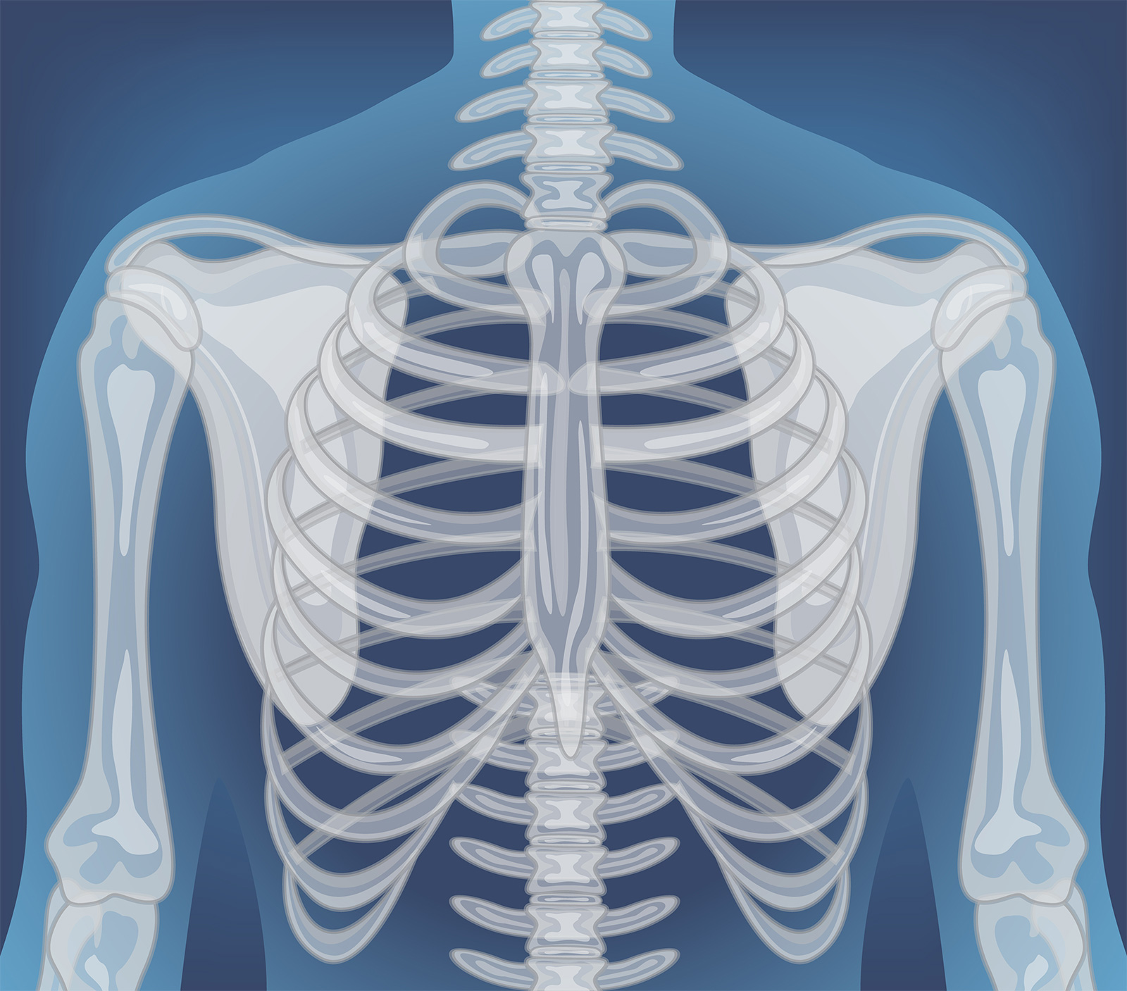
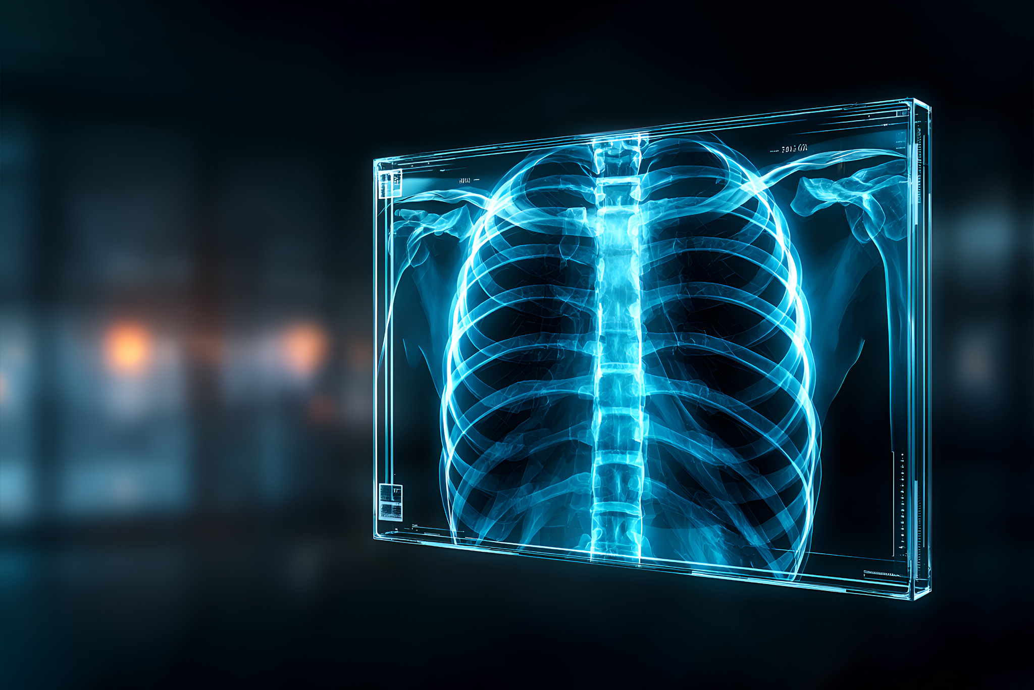
Digital Chest X-Ray (PA View)
The Chest PA (Posteroanterior) view is the most common diagnostic imaging test. At SR Scanning Center, we use advanced digital detectors that capture high-resolution images of the heart, lungs, airways, blood vessels, and the bones of the spine and chest.
This scan is essential for diagnosing pneumonia, lung cancer, tuberculosis (TB), and heart failure (cardiomegaly). The digital nature of our X-ray allows us to manipulate the image brightness and contrast instantly, revealing subtle details in the lung fields that might be missed on conventional film.
It is also the first line of investigation for chest pain, chronic cough, or shortness of breath. The procedure is extremely fast, taking only a fraction of a second, with results available immediately for your doctor.
Key Highlights
- Crystal clear imaging of Lungs and Heart.
- Screening for Tuberculosis and Pneumonia.
- Evaluates Rib Fractures and Chest Trauma.
- Instant digital reporting.
Spine X-Ray (Cervical / Dorsal / Lumbar)
Back and neck pain are common complaints. Our Digital X-ray services provide detailed views of the vertebral column. We perform Cervical (Neck), Dorsal (Upper Back), and Lumbar/Lumbosacral (Lower Back) spine X-rays to assess bone alignment and integrity.
These X-rays are crucial for detecting Spondylosis (wear and tear), fractures from trauma, and Spondylolisthesis (slipped vertebrae). We routinely perform flexion and extension views to check for spinal instability which static images might miss.
It is an excellent initial screening tool before proceeding to more expensive tests like MRI. Our digital system ensures that even fine hairline fractures are visible.
Key Highlights
- Diagnosis of Spondylosis and Arthritis.
- Evaluation of Traumatic Spinal Fractures.
- Assessment of Kyphosis and Scoliosis.
- Functional views (Flexion/Extension) available.
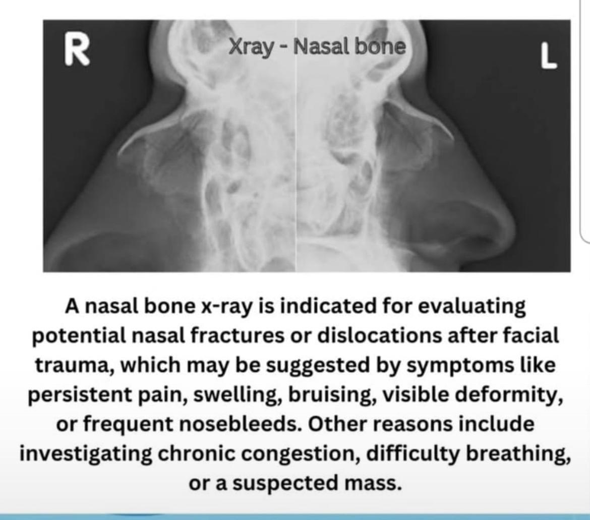
PNS (Waters View) & Skull X-Ray
The PNS (Paranasal Sinuses) Waters view is a specialized X-ray used to diagnose sinusitis. It clearly visualizes the maxillary sinuses to check for fluid levels, mucosal thickening, or polyps that cause blockage and headaches.
Skull X-rays are primarily used to assess fractures of the calvarium (skull bone) following head trauma. They are also used to detect congenital anomalies or bone diseases affecting the cranium.
This is a quick, non-invasive way to evaluate facial pain, nasal congestion, or head injuries.
Key Highlights
- Rapid diagnosis of Sinusitis.
- Detection of Skull Fractures.
- Evaluation of Nasal Bone fractures.
- Adenoid X-rays for pediatric breathing issues.
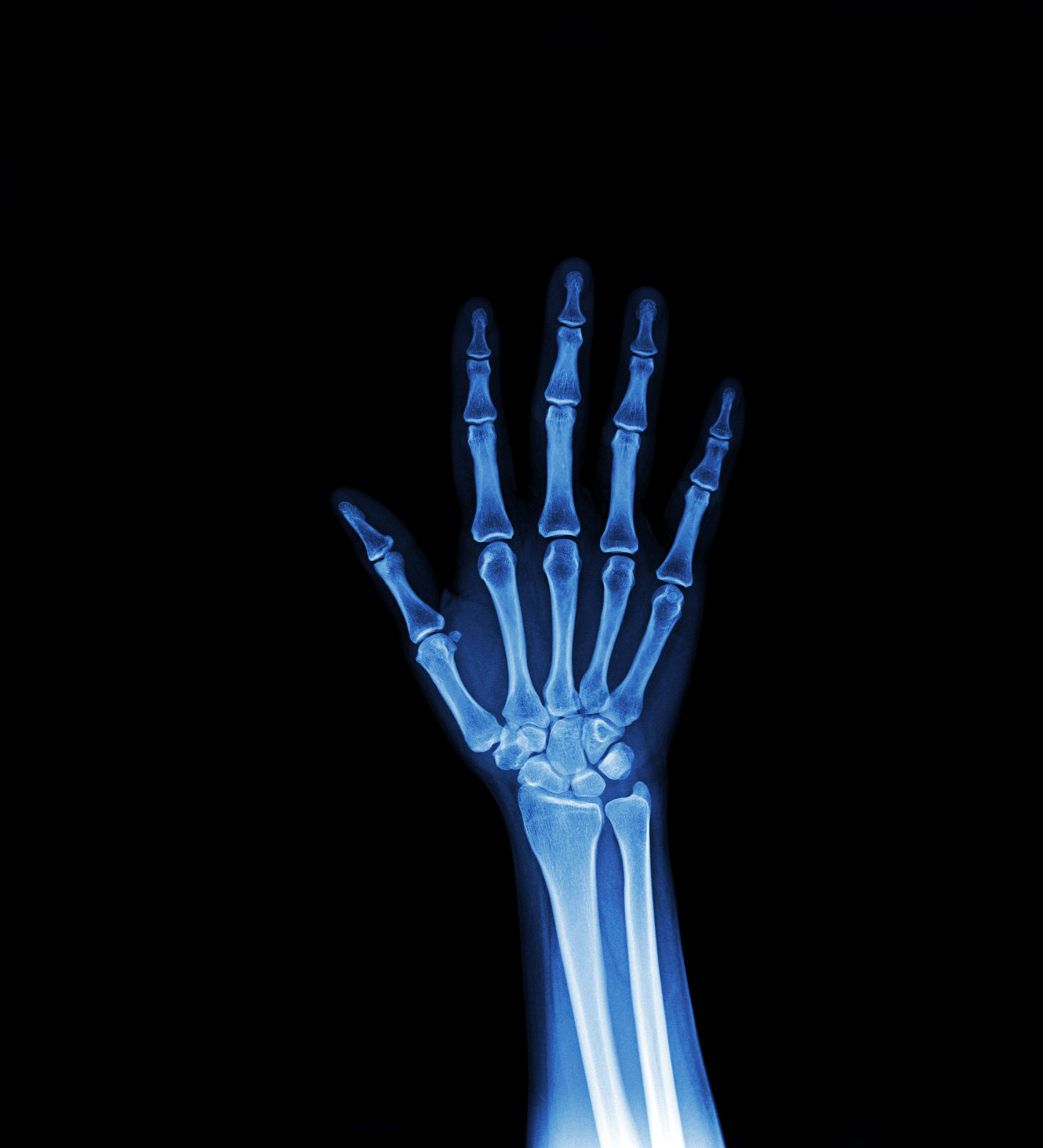
Extremities (Hand, Wrist, Leg, Foot)
From sports injuries to accidental falls, X-rays of the extremities are vital for orthopedics. We provide high-quality AP (Anteroposterior) and Lateral views for hands, wrists, arms, legs, and feet.
Our digital sensors provide exceptional detail of the trabecular bone pattern, allowing for the detection of fractures, dislocations, and bone infections (osteomyelitis). We also assess bone age in children to monitor growth disorders.
Whether it is a "Left hand AP View" or a complex foot fracture, our technicians ensure precise positioning for the most diagnostic image.
Key Highlights
- Immediate fracture detection.
- Assessment of Joint Dislocations.
- Bone Age estimation for Pediatrics.
- Evaluation of foreign bodies (glass/metal).
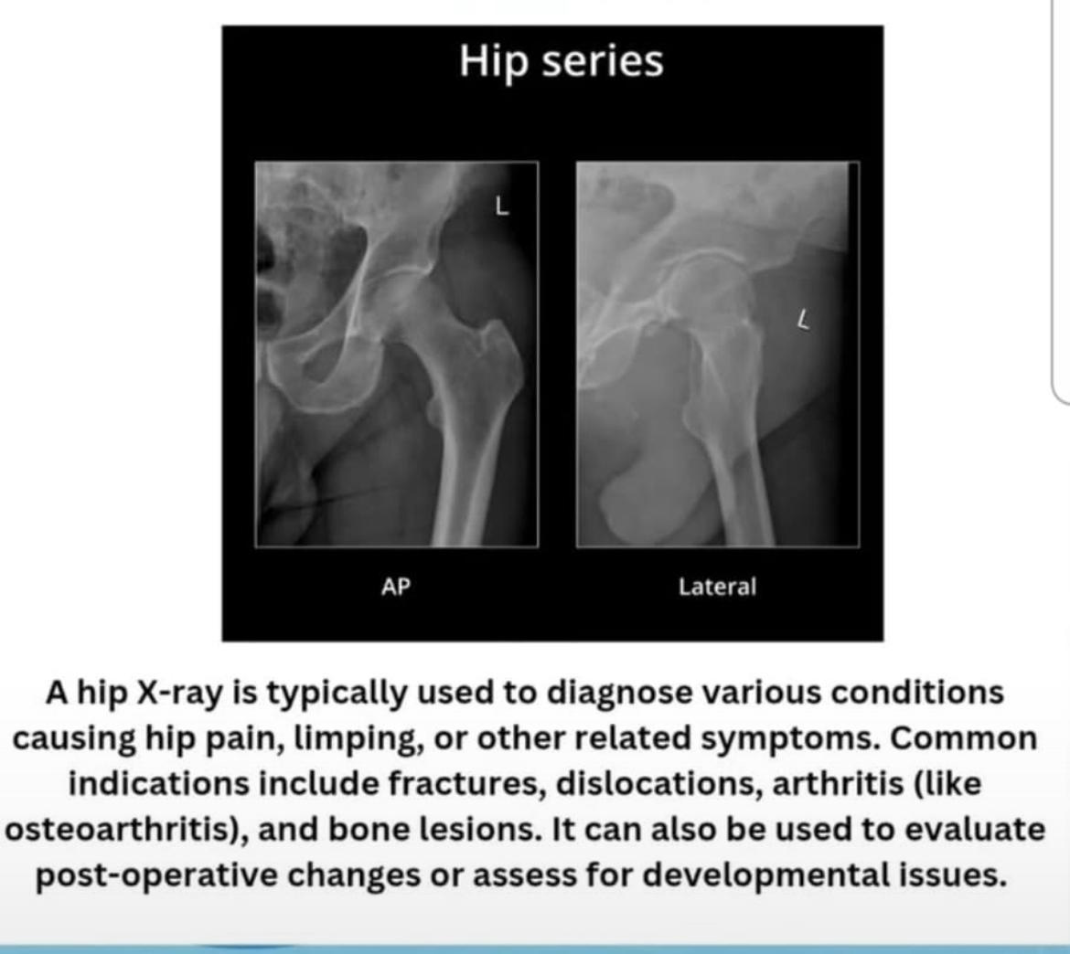
Abdomen & Pelvis X-Ray
Abdominal X-rays (KUB) are frequently used to detect kidney stones (Renal Calculi). Since many stones are radiopaque (contain calcium), they show up clearly on our digital X-rays. We perform both Supine (lying down) and Erect (standing) views.
The Erect Abdomen view is critical for diagnosing intestinal obstruction or perforation (free air under the diaphragm), which are surgical emergencies.
Pelvis X-rays are standard for diagnosing hip fractures in the elderly and screening for hip dysplasia in infants. We also evaluate the sacroiliac joints for ankylosing spondylitis.
Key Highlights
- Detection of Kidney and Bladder Stones.
- Diagnosis of Bowel Obstruction.
- Evaluation of Hip Fractures and Arthritis.
- Emergency assessment for perforation.
Joint X-Rays (Knee, Shoulder, Hip)
Chronic joint pain requires a detailed look at the bone spacing and surface. Our Digital X-ray is the gold standard for diagnosing Osteoarthritis (OA) and Rheumatoid Arthritis (RA). It reveals narrowing of the joint space, bone spurs (osteophytes), and erosions.
For shoulder pain, we can identify calcific tendinitis or dislocation. For knee pain, standing weight-bearing views help the orthopedic surgeon determine the severity of arthritis and the need for knee replacement surgery.
Key Highlights
- Gold standard for Osteoarthritis grading.
- Detects loose bodies in joints.
- Evaluates alignment for joint replacement planning.
- Post-operative checks for implants.
Whole Spine & Long Leg (Image Stitching)
SR Scanning Center is equipped with advanced Image Stitching Software. Standard X-ray plates are not long enough to capture the entire spine or the entire leg in one go. Image stitching solves this by combining multiple images into one seamless panoramic view.
Whole Spine Scanography: Essential for diagnosing and measuring Scoliosis (curvature of the spine) and Kyphosis. It allows the doctor to measure the Cobb angle accurately for treatment planning.
Long Leg Scanogram: Used to measure Limb Length Discrepancy (difference in leg lengths) and mechanical axis alignment. This is vital for planning corrective osteotomy surgeries and total knee replacements to ensure perfect alignment.
Key Highlights
- Full-length imaging of Spine (Scoliosis Study).
- Full-length imaging of Legs (Scanogram).
- Seamless digital stitching for accurate measurement.
- Essential for orthopedic surgical planning.
FAQs - Digital X-Ray
Yes. Digital X-ray technology requires significantly less radiation dose compared to older film-based X-rays. The exposure is extremely low and considered safe for diagnostic purposes.
You must inform the technician if there is any chance you are pregnant. X-rays are generally avoided during pregnancy unless absolutely necessary. Ultrasound is the preferred method for pregnant patients.
Yes. Metal objects like necklaces, zippers, buttons, and bras with underwire can block X-rays and create artifacts on the image. You may be asked to change into a hospital gown.

Supporting Our Communities with Safe & Crystal Clear Imaging.
At SR Scanning Center, we utilize high-frequency Digital X-Ray systems to ensure minimal radiation exposure for our patients. Our commitment to safety means we provide diagnostic accuracy without compromising your long-term health.
With over 90% of health decisions based on diagnostic results, we are committed to being a trusted healthcare partner you need for critical decisions.
Quick & Comfortable
Digital sensors capture images instantly, significantly reducing wait times and the need for retakes due to patient movement.
High Def Imaging
Superior contrast allows for better visualization of fine fractures, kidney stones, and soft tissue shadows compared to traditional film.
Process
Preparing for your X-Ray is Simple and Quick.
To reduce your wait time, simple preparation is key. Most routine X-rays require no special fasting (except for some abdominal studies). The most important step is ensuring you are not wearing metal objects in the area being scanned.
Our digital system connects directly with your doctor to deliver results instantly, ensuring you get the care you need without delay.
-
Walk-ins Welcome
-
Instant Digital Prints
-
Lowest Radiation Dose
Visit Us
Bring your doctor's prescription. For most X-rays, no prior appointment is needed, and wait times are minimal.
Contact UsPreparation
Remove jewelry, belts, or clothing with metal zippers. Inform us immediately if there is a chance of pregnancy.
Book a VisitThe Scan
Our technician will position you comfortably. The actual X-ray exposure takes less than a second and is painless.
Services InfoGet Results
You will receive high-quality digital films and a report to share with your consulting doctor immediately.
View Results